How Ultrasound Works
by Craig C. Freudenrich, Ph.D. There are many situations in which ultrasound is performed. Perhaps you are pregnant, and your obstetrician wants you to have an ultrasound to check on the developing baby or determine the due date. Maybe you are having problems with blood circulation in a limb or your heart , and your doctor has requested a Doppler ultrasound to look at the blood flow. Ultrasound has been a popular medical imaging technique for many years.
Ultrasound examination during pregnancy
In this edition of How Stuff Works , we will look at how ultrasound works, what type of ultrasound techniques are available and what each technique can be used for.
What is Ultrasound?
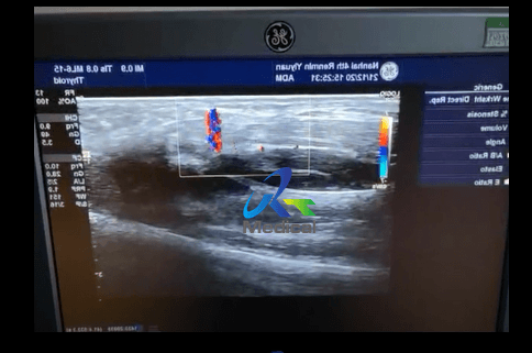
Ultrasound or ultrasonography is a medical imaging technique that uses high frequency sound waves and their echoes. The technique is similar to the echolocation used by bats, whales and dolphins, as well as SONAR used by submarines . In ultrasound, the following events happen: The ultrasound machine transmits high-frequency (1 to 5 megahertz) sound pulses into your body using a probe. The sound waves travel into your body and hit a boundary between tissues (e.g. between fluid and soft tissue, soft tissue and bone). Some of the sound waves get reflected back to the probe, while some travel on further until they reach another boundary and get reflected. The reflected waves are picked up by the probe and relayed to the machine. The machine calculates the distance from the probe to the tissue or organ (boundaries) using the speed of sound in tissue (5,005 ft/s or1,540 m/s) and the time of the each echo's return (usually on the order of millionths of a second). The machine displays the distances and intensities of the echoes on the screen, forming a two dimensional image like the one shown below.
Photo courtesy Karim and Nancy Nice
Ultrasound image of a growing fetus (approximately 12 weeks old) inside a mother's uterus. This is a side view of the baby, showing (right to left) the head, neck, torso and legs. In a typical ultrasound, millions of pulses and echoes are sent and received each second. The probe can be moved along the surface of the body and angled to obtain various views.
A basic ultrasound machine has the following parts
transducer probe - probe that sends and receives the sound waves central processing unit (CPU) - computer that does all of the calculations and contains the electrical power supplies for itself and the transducer probe transducer pulse controls - changes the amplitude, frequency and duration of the pulses emitted from the transducer probe display - displays the image from the ultrasound data processed by the CPU keyboard/cursor - inputs data and takes measurements from the display disk storage device (hard, floppy, CD) - stores the acquired images printer - prints the image from the displayed data
Transducer Probe
The transducer probe is the main part of the ultrasound machine. The transducer probe makes the sound waves and receives the echoes. It is, so to speak, the mouth and ears of the ultrasound machine. The transducer probe generates and receives sound waves using a principle called the piezoelectric (pressure electricity) effect , which was discovered by Pierre and Jacques Curie in 1880. In the probe, there are one or more quartz crystals called piezoelectric crystals . When an electric current is applied to these crystals, they change shape rapidly. The rapid shape changes, or vibrations, of the crystals produce sound waves that travel outward. Conversely, when sound or pressure waves hit the crystals, they emit electrical currents. Therefore, the same crystals can be used to send and receive sound waves. The probe also has a sound absorbing substance to eliminate back reflections from the probe itself, and an acoustic lens to help focus the emitted sound waves. Transducer probes come in many shapes and sizes, as shown in the photo above. The shape of the probe determines its field of view, and the frequency of emitted sound waves determines how deep the sound waves penetrate and the resolution of the image. Transducer probes may contain one or more crystal elements; in multiple-element probes, each crystal has its own circuit. Multiple-element probes have the advantage that the ultrasounc beam can be "steered" by changing the timing in which each element gets pulsed; steering the beam is especially important for cardiac ultrasound (see Basic Principles of Ultrasound for details on transducers). In addition to probes that can be moved across the surface of the body, some probes are designed to be inserted through various openings of the body (vagina, rectum, esophagus) so that they can get closer to the organ being examined (uterus, prostate gland, stomach); getting closer to the organ can allow for more detailed views.
Central Processing Unit (CPU)
The CPU is the brain of the ultrasound machine. The CPU is basically a computer that contains the microprocessor , memory , amplifiers and power supplies for the microprocessor and transducer probe. The CPU sends electrical currents to the transducer probe to emit sound waves, and also receives the electrical pulses from the probes that were created from the returning echoes. The CPU does all of the calculations involved in processing the data. Once the raw data are processed, the CPU forms the image on the monitor. The CPU can also store the processed data and/or image on disk.