
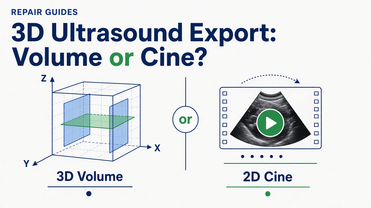
- What Did the Scanner Actually Export: A Volume, a Derived Image, or a Screenshot?
- Which DICOM SOP Class and Metadata Identify a True 3D Ultrasound Volume?
- Why Does a 3D Export Appear as Separate Slices or a Rotating Clip?
- How Do You Check Whether Both the Scanner and PACS Support a True Volume?
- When Is a Rendered Cine Acceptable—and When Is It the Wrong Deliverable?
- How Do You Separate an Export Limitation From a Probe, Motor Board, or Matrix Controller Fault?
- Synthetic Acceptance Test: Capture Object, Geometry, and Receiver Evidence Before Escalating
- Where Rongtao Medical Fits—and Where It Does Not
- Frequently Asked Questions
What Did the Scanner Actually Export: A Volume, a Derived Image, or a Screenshot?
For hospital healthcare technology management (HTM) engineers, independent service organizations (ISOs), and buyers accepting a used 3D/4D ultrasound system, few complaints create more misplaced troubleshooting than this ticket: "The 3D scan looks fine on the cart, but PACS only shows a rotating movie or flat slices—we cannot reslice it." Because a spinning 3D rendering on screen visually resembles a volume, teams often assume either the receiving workstation broke the file or the ultrasound system's 4D hardware is failing.
In DICOM, however, visual appearance on the console does not define what left the network port. Official DICOM guidance in PS3.17 Section PP.3 and normative definitions in PS3.3 Section A.59 divide 3D ultrasound exchange into two fundamentally separate categories: 3D volume data sets and 2D images derived from the volume. 1 3 On top of those standard classes, commercial scanners frequently emit screen-capture objects or store proprietary 3D/4D payloads inside private attributes of legacy multi-frame objects. 7 10
| DICOM SOP Class | SOP Class UID | What It Actually Carries | Supports Arbitrary Downstream MPR? | Common Acceptance Trap |
|---|---|---|---|---|
| Enhanced US Volume Storage | 1.2.840.10008.5.1.4.1.1.6.2 | Cartesian 3D volume or temporally related 3D+t volumes arranged as equally spaced parallel planes with per-frame geometry. | Designed for it, provided the receiving PACS or workstation supports the SOP Class and MPR reconstruction. | Assuming a conformant volume file proves every PACS can display it, or proves geometric accuracy of the scanner's scan-conversion. |
| Ultrasound Multi-frame Image Storage | 1.2.840.10008.5.1.4.1.1.3.1 | Multi-frame 2D loop: either a rendered 3D cine, pre-extracted MPR slices, spatially related source frames, or (in Standard Extended implementations) private 3D/4D tags. | Not in standard generic form, unless the file carries vendor-extended/private 3D metadata understood by a matching workstation. | Mistaking a spinning 3D-rendered loop for an interactive volume, or seeing separate slices when a generic viewer ignores private tags. |
| Ultrasound Image Storage | 1.2.840.10008.5.1.4.1.1.6.1 | Single-frame 2D ultrasound image—such as one frozen MPR plane, one rendered still, or one source slice. | No. A single 2D frame cannot reconstruct an arbitrary 3D volume. | Receiving individual 2D captures from a 3D exam and assuming the underlying volume series was archived. |
| Secondary Capture Image Storage (and Multi-frame True Color SC) | 1.2.840.10008.5.1.4.1.1.7 (and .7.4) | Display screenshot or screen-capture movie converted into a generic DICOM image wrapper. | No. Lacks volumetric geometry and ultrasound calibration structures. | Treating a screen-grabbed rotating 3D display as volumetric data—or even as calibrated 2D pixel geometry. |
Source: SOP Class names and UIDs from DICOM PS3.4 Table B.5-1 (2026d); object semantics from PS3.17 PP.3/PP.3.2, PS3.3 A.59, and SlicerHeart technical documentation. See sources 1, 2, 3, 6, and 10.
Before touching a probe connector, opening a rear panel, or blaming a PACS gateway, inspect the Service-Object Pair (SOP) Class Unique Identifier (UID) of the received instance. That single tag tells you immediately whether the receiving archive holds a standardized Cartesian volume, a 2D derived object, or a screen capture.
Which DICOM SOP Class and Metadata Identify a True 3D Ultrasound Volume?
Under DICOM PS3.4 Table B.5-1 and PS3.3 Section A.59, the standardized object built for interchangeable 3D and 4D ultrasound volumes is Enhanced US Volume Storage (SOP Class UID 1.2.840.10008.5.1.4.1.1.6.2). 3 6 Unlike conventional ultrasound multi-frame loops—which evolved for 2D temporal cine playback—Enhanced US Volume uses the enhanced multi-frame architecture with shared and per-frame functional groups so any compliant receiver knows where each plane sits in 3D space and time. 1
To distinguish a true standard volume from an ordinary multi-frame series during acceptance testing, verify five normative markers defined in PS3.3 Section A.59.4 and Section C.8.24.3: 3 4
- Volumetric identity constraints: In the US Image Description Macro,
Volumetric Properties (0008,9206)must equalVOLUMEandVolume Based Calculation Technique (0008,9207)must equalNONE. 3 - Dimension organization: In the Enhanced US Image Module,
Dimension Organization Type (0020,9311)is enumerated as either3D(a single static volume) or3D_TEMPORAL(a time series of volumes, such as 4D or Spatial-Temporal Image Correlation, STIC). Even for a static 3D volume,Dimension Index Sequence (0020,9222)must contain exactly three items: (1) the temporal position attribute, (2)Image Position (Volume) (0020,9301)insidePlane Position (Volume) Sequence (0020,930E), and (3)Data Type (0018,9808)insideImage Data Type Sequence (0018,9807)(associating tissue, flow, or other planes at the same position and time). 1 4 - Cartesian plane geometry: Each Cartesian volume consists of parallel planes with equal spacing in the Z direction.
Plane Position (Volume)is mandatory per frame (not shared), with X and Y values ofImage Position (Volume) (0020,9301)fixed to0. Conversely,Plane Orientation (Volume)is mandatory as a shared functional group (not per-frame), withImage Orientation (Volume) (0020,9302)constrained to1\0\0\0\1\0. 3 - Acquisition geometry link (when patient-referenced): If
Ultrasound Acquisition Geometry (0020,9307)equalsPATIENT, the object must also includePlane Position (Patient)andPlane Orientation (Patient)functional group macros. 3 - Position measuring method:
Position Measuring Device Used (0018,980C)records how spatial positioning was obtained—RIGID(rigidly connected mechanical/fixed geometry),TRACKED(tracked freehand), orFREEHAND(untracked freehand)—signaling to downstream users how reliable voxel and inter-plane spacing assumptions may be. 4
Notice a crucial boundary written directly into both PS3.17 PP.3 and PS3.3 A.59.1: the DICOM standard explicitly places the method used to generate the Cartesian volume, its mathematical relationship to non-Cartesian acquisition frames, and the algorithms used for MPR or 3D rendering outside the standard's scope. 1 3 A valid Enhanced US Volume file guarantees standardized structural encoding; it does not by itself prove that a scanner's scan-conversion was accurate or that two different PACS viewers will render identical 3D surfaces.
Why Does a 3D Export Appear as Separate Slices or a Rotating Clip?
When an operator exports a 3D acquisition and sees a flat cine loop or a stack of unlinked 2D slices at the destination, one of four documented software or standards mechanisms is usually responsible—none of which implies a broken ultrasound transducer.
- The scanner intentionally exported 2D derived images. PS3.17 Section PP.3.2 explains that 3D render views (projections), separate MPR views, and sets of spatially related source frames are encoded using the standard
Ultrasound ImageorUltrasound Multi-frame ImageIODs for backward compatibility with routine review viewers. In these objects,Image Type (0008,0008)Value 4 uses a hexadecimal modality bit map defined in PS3.3 C.8.5.6.1.1 where0040flags 3D Rendering and0400flags Spatially-related Frames (combined additively with other active modes—for example,0041for 2D Imaging plus 3D Rendering). Optionally,Source Image Sequence (0008,2112)andDerivation Code Sequence (0008,9215)link back to the source volume for two-stage review. 2 5 - The scanner stores 3D/4D inside Standard Extended or private tags that generic viewers cannot interpret. The SlicerHeart project's import documentation—which describes reader paths for GE, Philips, Samsung, and other scanners—reports that most ultrasound manufacturers do not store 3D/4D volumes in standard DICOM fields; standard fields often hold only 2D screenshots or screen-capture videos, while true volumetric data resides in private attributes requiring vendor-supplied libraries or proprietary workstations. 10 Philips' official EPIQ and Affiniti Release 11.0.x DICOM Conformance Statement provides a concrete, release-specific example: it does not list Enhanced US Volume Storage, instead using Standard Extended
Ultrasound Multi-frame Image StoragewherePresentation Intent Type (0008,0068) = "FOR PRESENTATION"signals spatially related 3D frames, while 3D Temporal (4D) volume data is placed in private attributes whose transmission depends on per-destination archive/media advanced settings. 7 - The receiving software expects standard geometry that legacy multi-frame objects lack. As DICOM expert David Clunie observes in commentary quoted by SlicerHeart, the older Ultrasound Multi-frame Image IOD does not encode 3D volume geometry, because it dates from the video-capture era. 10 In a 3D Slicer community thread, a researcher who wrote a reconstructed ultrasound volume out as US DICOM from research software (not from a clinical scanner) saw it reload as many separate slices, each with a spacing-calibration warning, rather than one coherent volume. The replies noted that there is no commonly used DICOM format for 3D/4D ultrasound and asked which DICOM information objects the reading software actually supports. The same receiver behavior can greet a scanner export, which is why the receiving side must be checked before the sender is blamed. 11
- Workstation or export-preset filtering dropped or bypassed the volume. A site can configure the scanner or an intermediate router to send only derived rendered loops or Secondary Capture snapshots to the main PACS, for storage or universal-display reasons, while the volume data stays on the console or goes only to a dedicated 3D review workstation. Converting a proprietary volume afterwards is not a clean fallback either: SlicerHeart documents that the Philips QLAB "Cartesian DICOM" export option writes volumes into standard fields but omits required identifiers such as SOP Instance UID, Study/Series Instance UIDs, and patient name and ID, so the files must be patched before a standard DICOM database will ingest them. 10
How Do You Check Whether Both the Scanner and PACS Support a True Volume?
Interoperability requires a match on both sides of the DICOM association: the ultrasound console (acting as Storage SCU, or Service Class User) must be configured and licensed to generate the object, and the PACS, vendor-neutral archive (VNA), or workstation (acting as Storage SCP, or Service Class Provider) must accept that SOP Class UID in presentation-context negotiation—and have an application module able to reconstruct it. 9 11
Table 2 illustrates how sharply release-specific conformance declarations differ across real ultrasound systems and receivers—from full-featured carts (Philips EPIQ and Affiniti Release 11.0.x) to handheld scanners (Philips Lumify R5.0) and enterprise PACS viewers (Visage 7 Version 12.00). 7 8 9 Never assume a product family supports Enhanced US Volume simply because it performs 3D or 4D scanning on the console, and never generalize a single software revision's table to older or newer releases.
| Product & Document Revision | Role | Enhanced US Volume Storage (1.2.840.10008.5.1.4.1.1.6.2) | How 3D/4D or Derived Data Is Declared | Practical Verification Note |
|---|---|---|---|---|
| Philips EPIQ & Affiniti Family, Release 11.0.x (D001649514 Rev A, 15 Apr 2024) | Modality (SCU / SCP) | Not listed in Storage SOP Class table (Section 4.2.1.1, p. 24). | Standard Extended US / US Multi-frame Storage: Presentation Intent Type (0008,0068) = FOR PRESENTATION for 3D spatially related frames; private attributes for 4D; optional Private 3D Presentation State (1.3.46.670589.2.5.1.1) on selected EPIQ models. | Check per-destination Archive / Media > Advanced private-attribute export flags; a generic PACS may archive the frames without reconstructing the private 4D structure. |
| Philips Lumify R5.0 (HSDP-1299212 v01, 17 Aug 2023) | Handheld Ultrasound (SCU) | Not listed. | US Image Storage (.6.1), US Multi-frame Image Storage (.3.1), and Secondary Capture (.7) only. | A 2D handheld, included as a contrast: its declared objects are 2D stills, 2D multi-frame cine loops, and screen captures, not Cartesian 3D volume objects. |
| Visage 7 / Visage Ease Pro, Version 12.00 (18 Jun 2026) | PACS / Viewer (SCU & SCP) | Listed as Storage SCU (p. 23), Storage SCP (p. 43), and Viewable (X) (added in Version 07.00, 11 May 2016). | Accepts and views Enhanced US Volume alongside US Multi-frame, US Image, and Secondary Capture. | Even when a PACS supports Enhanced US Volume SCP, a scanner that exports private multi-frame tags or 2D rendered clips will still display only what the scanner actually sent. |
Source: Directly inspected from release-specific vendor DICOM Conformance Statements. These rows document stated capabilities for the named software revisions only and do not certify other releases or live site compatibility. See sources 7, 8, and 9.
When auditing a mismatch, pull the exact software version from the ultrasound system and the exact build from the PACS or workstation, then compare their Storage SOP Class tables side by side. Remember that conformance statements are manufacturer self-declarations; an active software option, a specific destination profile, or a transfer-syntax restriction can still prevent volume transmission on an installed unit even when the PDF lists the SOP Class.
When Is a Rendered Cine Acceptable—and When Is It the Wrong Deliverable?
An important counter-evidence rule keeps teams from manufacturing false defects: receiving a 2D rendered cine instead of an Enhanced US Volume object is not automatically an error, data loss, or hardware malfunction. DICOM PS3.17 Section PP.3.2 explicitly accommodates a "Two-Stage Review" workflow in which a reviewing clinician inspects pre-rendered 3D projections or representative MPR slices in a routine PACS viewer, while links in Source Image Sequence and Referenced Image Sequence preserve traceability back to the source volume when deeper interrogation is needed. 2
- When a rendered cine or MPR slice set is acceptable: Routine documentation of a surface-rendered fetal, cardiac, or pelvic view where the department only reviews pre-selected angles or a fixed fly-through loop in PACS, and either no downstream re-slicing is required or the raw volume remains accessible on the console or a dedicated vendor workstation.
- When a rendered cine is the wrong deliverable: Quantitative volumetric re-analysis on a third-party workstation, arbitrary off-axis MPR slicing after the patient leaves, research repositories requiring standardized Cartesian geometry, or pre-purchase acceptance of a used 3D/4D system where the buyer specifically contracted for PACS-archived volumetric export.
If your team intentionally uses a rendered multi-frame cine (1.2.840.10008.5.1.4.1.1.3.1) and discovers a different problem—such as the rotating loop playing at double speed or stuttering—do not confuse playback timing with volume geometry. Inspect the frame intervals and viewer defaults using our ultrasound cine frame timing guide. Similarly, if volumetric measurements visible on the console are absent in the reporting system, follow the DICOM SR measurement delivery guide rather than treating measurements as part of pixel export.
How Do You Separate an Export Limitation From a Probe, Motor Board, or Matrix Controller Fault?
Hardware repair teams routinely receive unnecessary 4D probe or board inquiries triggered purely by PACS display complaints. The isolation gate is straightforward: verify acquisition and local reconstruction on the ultrasound console itself before evaluating export.
| Observed Behavior | Most Likely Domain | First Verification Action | Escalation / Next Route |
|---|---|---|---|
| 3D/4D sweep and MPR reslicing work normally on the console; PACS shows a rotating movie or flat slices. | Export configuration, SOP class choice, or PACS/viewer support. Working local acquisition is not evidence of a probe or board fault. | Inspect exported SOP Class UID, Image Type value 4, private-tag destination flags, and both conformance statements. | PACS administrator, clinical applications specialist, or OEM DICOM integration team. |
| Console acquires 3D/4D locally, but nothing transfers to PACS (association fails, port dead, or timeouts across all exam types). | Network/AE configuration, TLS/clock, or rear I/O / host hardware. | Separate protocol rejection from physical Ethernet/I/O failure using a synthetic test send. | Follow the DICOM network and I/O board fault guide. |
| Console acquires 2D normally, but 3D/4D sweep stalls, grinds, leaks oil, throws a motor error, or fails to build any volume locally. | Acquisition hardware: mechanical volume probe, console 4D motor drive board, or electronic matrix controller. | Test a second known-good volume probe of the same class; inspect dome/cable safely without pressing the lens. | Follow the 4D volume acquisition hardware decision path or request probe repair. |
| Buying a used 3D/4D cart and verifying whether volume export meets clinical or workstation requirements. | Pre-purchase acceptance and license/option verification. | Run a synthetic phantom volume export to the target PACS/workstation during Gate 2/3 acceptance. | Combine this test with the used ultrasound system acceptance guide. |
Source: Engineering synthesis combining DICOM PS3.17/PS3.3/PS3.4 rules with Rongtao hardware fault-isolation boundaries. See sources 1, 2, 6, 7, 11, and 12.
Never order a replacement 4D motor board, matrix controller, or volume transducer solely because an exported DICOM file opened as a 2D cine in PACS. Conversely, if local console 3D/4D acquisition itself fails—for example, 2D B-mode works cleanly while volume sweep throws motor-control errors—an export setting will not restore physical sweep motion, and safe hardware isolation is required.
Synthetic Acceptance Test: Capture Object, Geometry, and Receiver Evidence Before Escalating
Use a non-patient phantom or synthetic test scan—never upload patient-identifiable clinical DICOM files to public tools or external service desks—and run these six checks inside facility-approved inspection tools before raising a software or hardware ticket:
- Confirm local console 3D/4D reconstruction first. Acquire a synthetic volume sweep on a tissue-mimicking phantom and verify that A/B/C MPR planes and volume rendering rotate and reslice interactively on the scanner console.
- Export to the target destination and inspect the SOP Class UID
(0008,0016). Record whether the archived instance is1.2.840.10008.5.1.4.1.1.6.2(Enhanced US Volume),1.2.840.10008.5.1.4.1.1.3.1(US Multi-frame),1.2.840.10008.5.1.4.1.1.6.1(US Image), or1.2.840.10008.5.1.4.1.1.7(Secondary Capture). 6 - Check volume geometry versus derived-image tags. For Enhanced US Volume, confirm
Volumetric Properties (0008,9206) = VOLUME,Dimension Organization Type (0020,9311)(3Dor3D_TEMPORAL), three items inDimension Index Sequence (0020,9222), and per-frameImage Position (Volume) (0020,9301). For US / US Multi-frame, inspectImage Type (0008,0008)Value 4 (such as0040or0400bits),Presentation Intent Type (0008,0068), and anySource Image Sequence (0008,2112)orDerivation Code Sequence (0008,9215)links. 2 3 4 5 - Audit console destination presets and private-tag handling. Check the scanner's DICOM archive destination profile (for example, whether "3D/Volume," "Normal 2D," or private-tag export flags are enabled, and whether an intermediate DICOM router strips private attributes).
- Verify the receiver's DICOM Conformance Statement and MPR license. Confirm that the receiving PACS or workstation explicitly lists Enhanced US Volume Storage as SCP (if standard volumes are expected) or supports the OEM's specific extended/private volume format.
- Document the exact acceptance verdict. Record the console software build, destination AE profile, received SOP Class UID, frame count, and whether interactive MPR succeeded on the target viewer—preserving the synthetic test files for vendor escalation.
The comparison below shows what a synthetic inspection summary looks like across the two most common outcomes. These values are constructed strictly for structural illustration and do not represent a real patient scan or a specific OEM software dump.
Outcome A — Standardized 3D Volume (Synthetic Subset)SOP Class UID (0008,0016): 1.2.840.10008.5.1.4.1.1.6.2 (Enhanced US Volume Storage)
Volumetric Properties (0008,9206): VOLUME
Volume Based Calculation Technique (0008,9207): NONE
Dimension Organization Type (0020,9311): 3D
Image Orientation (Volume) (0020,9302): 1\0\0\0\1\0
Image Position (Volume) (0020,9301): 0\0\z_k (per-frame, uniform Z step)
Position Measuring Device Used (0018,980C): RIGID
Outcome B — 2D Derived Rotating Cine (Synthetic Subset)SOP Class UID (0008,0016): 1.2.840.10008.5.1.4.1.1.3.1 (Ultrasound Multi-frame Image Storage)
Image Type (0008,0008): DERIVED\SECONDARY\ABDOMINAL\0041 (0001 2D + 0040 3D Rendering)
Source Image Sequence (0008,2112): References source volume SOP Instance UID (if linked)
Derivation Code Sequence (0008,9215): CID 7203 Image Derivation code
Where Rongtao Medical Fits—and Where It Does Not
Rongtao Medical is an independent multi-brand ultrasound repair and replacement-parts provider—not a PACS software developer, DICOM gateway vendor, or OEM-authorized representative. If your synthetic acceptance test shows that the console sweeps and reconstructs 3D/4D cleanly on the cart and only the exported DICOM object is a 2D cine or private-tag mismatch, the fix belongs to your PACS administrator, applications team, or OEM DICOM configuration support—not a parts purchase.
Where Rongtao Medical directly helps is when Step 1 of the isolation test reveals a genuine acquisition or I/O hardware failure: a mechanical 4D volume probe with sweep-motor, encoder, or oil-bladder symptoms; a failed console 4D motor drive board or matrix controller; or a dead physical network/I/O interface. Rongtao supports component-level board repair backed by 48-hour real-machine testing inside live ultrasound platforms, a standard 5–8 business-day board-repair turnaround, and a typically 90-day warranty (exact terms confirmed per quote), alongside probe and transducer repair quoted per probe. Review our 10-step service process and quality verification standards for full documentation details. 12
When hardware isolation points to a failed board or volume probe, send a quote-ready package via Rongtao's contact desk containing:
- OEM, system model, and software revision (for example, GE Voluson E10 or Philips EPIQ 7; examples of the detail needed, not a list of supported systems).
- Manufacturer part number and clear photos of both sides of the board or probe label (only captured safely by qualified personnel; never open an energized cart).
- Exact symptom separation: confirm whether 2D B-mode works, whether 3D/4D fails locally on the console or only after DICOM export, and any exact on-screen error codes.
- Quantity and destination country so shipping, customs documentation, and lead-time terms can be confirmed accurately.
Frequently Asked Questions
Is an Enhanced US Volume object always required for 3D ultrasound?
No. DICOM PS3.17 defines both Enhanced US Volume (1.2.840.10008.5.1.4.1.1.6.2) for standardized Cartesian volumes and 2D derived objects (Ultrasound Image or Ultrasound Multi-frame Image) for pre-rendered clips, extracted MPR views, and spatially related frames. Many clinical scanners also rely on Standard Extended multi-frame objects or private formats paired with proprietary workstations. Whether Enhanced US Volume is required depends on your downstream receiver and workflow. 1 2 7 10
Can a PACS display a 3D volume if it only supports Ultrasound Multi-frame Image Storage?
Using standard DICOM fields alone, a viewer that only supports generic Ultrasound Multi-frame Image Storage (1.2.840.10008.5.1.4.1.1.3.1) will play the frames sequentially as a 2D cine loop or display individual slices—not reconstruct an arbitrary Cartesian volume—unless the file embeds vendor-specific extended or private 3D metadata and the viewer has a matching proprietary plugin or decoder. 10 11
Why do some 3D ultrasound files open only in the manufacturer's software?
Because many ultrasound manufacturers store raw 3D and 4D volumetric geometry inside private DICOM attributes or proprietary file wrappers while placing only 2D screenshots or rendered cine loops in standard public fields. Only a matching vendor workstation—or specialized decoding libraries and patchers designed for that OEM format—can parse those private fields into a 3D volume. 7 10
Does a 3D-looking image on the console prove the system exported a volume?
No. A system can acquire and render a 3D volume locally while its DICOM destination preset exports a 2D rendered clip (Image Type Value 4 bit 0040), a Secondary Capture screenshot (1.2.840.10008.5.1.4.1.1.7), or a multi-frame object stripped of private volumetric attributes. Always inspect the archived object's SOP Class UID and metadata at the receiver. 2 5 7
What should a buyer ask for when accepting a used 3D or 4D ultrasound system?
Beyond physical 2D and 4D sweep checks on the cart, run a synthetic phantom 3D export to your facility's actual PACS or review workstation before signing off. Require proof of the exact installed software revision, active DICOM/3D licenses, the exported SOP Class UID, and verification that the target receiver can perform whatever task the clinical workflow requires—whether that is interactive MPR or simple rendered-cine review. 9 11