
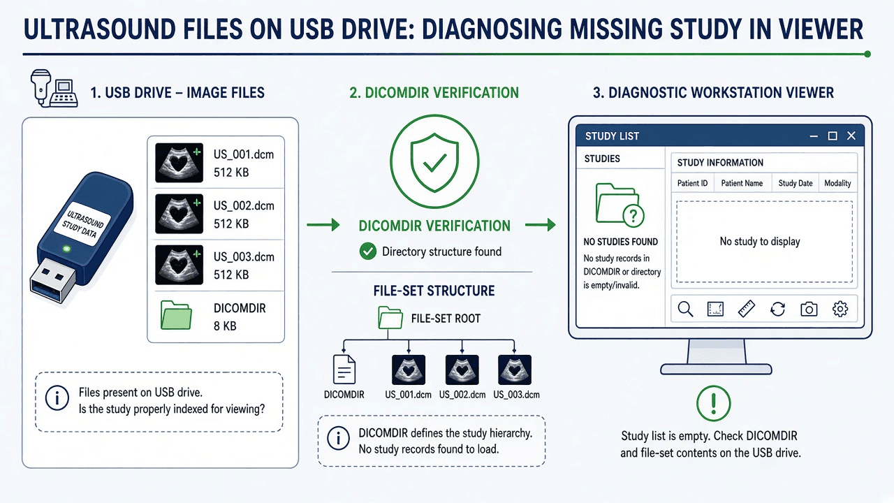
Why can files be present while the viewer lists no study?
This guide starts after files have reached removable media. A file manager may show occupied space and filenames, but it does not establish that the intended examination was completely exported, that each file is readable, or that the receiving software supports it. Start with observations rather than a presumed failed board: does the application find no patient at all, list a study whose images will not open, or display still images while omitting clips or reports? Those are different failure points.
A DICOM media directory is an index into a collection of files. A viewer using that index can fail to find objects when the index is absent or references paths that no longer exist. Another viewer may scan individual files and find the same objects without the directory. Keep the import method alongside the result; “opens on another computer” is useful evidence only when the application, version and action are known.
Record the original export selection, available export-completion message, media identifier and subsequent copy history. If the failure occurred before files reached the drive, use the guide to Sonosite PX USB export availability, whose model-specific administrator and study-state checks address an earlier stage. Network delivery is a separate route: query succeeds but C-MOVE retrieval fails concerns associations rather than USB directory structure.
What does DICOMDIR identify?
PS3.10 defines a File-set as files sharing a naming space and requires one file with the reserved File ID DICOMDIR in each File-set. PS3.10 also allows mappings in which a directory tree represents a File-set; “one per File-set” is the general rule, rather than a universal rule that every physical drive has exactly one. 1
For the STD-US ultrasound media profile, PS3.11 C.3.3 describes PATIENT, STUDY, SERIES and IMAGE directory records and requires references to the files incorporating objects defined for that profile. Its note places DICOMDIR at the medium root. Check the exporter’s declared profile and preserve its complete structure; do not assume every ultrasound export uses this particular profile. 4
File IDs contain one to eight components, each one to eight characters. Their structure carries no clinical meaning. A name that looks arbitrary is not evidence of damage, and an operating-system extension such as .dcm is not a reliable format test: alternate filenames can exist. Do not rename files merely to make them recognizable. 2
The ultrasound profile recommends random component names to reduce collisions, with existence checks when updating media. That recommendation explains a naming pattern; it does not establish what a particular scanner does when another export is already present. 5
| Layer | Evidence to collect | What it does not prove |
|---|---|---|
| Files on media | Original file/folder listing; read errors; transfer history | Occupied space or timestamps do not prove the intended exam is complete. |
| Directory index | DICOMDIR location and referenced files | A directory can contain metadata even when its target files are absent. |
| Object recognition | Actual file type, object class and encoding from approved inspection tools | A filename extension does not establish DICOM validity. |
| Viewer import and display | Product/version, command, loaded objects and error text | Successful display of one object does not prove every expected deliverable arrived. |
Source: Editorial evidence framework informed by sources 1–4; observations must be verified locally.
Which handling changes can break the directory references?
Work from the history of this export. Ask whether anyone copied only an image subfolder, renamed a file, moved DICOMDIR, reorganized folders or combined independent exports. These are possible failure mechanisms, not a measured ranking of causes. Keep the source media unchanged while an authorized person compares a controlled working copy with the original structure.
Partial copying: copying an image folder while leaving its directory behind can break a directory-based import. Conversely, copying only DICOMDIR can leave a plausible study list whose referenced files are missing. Copying the whole tree preserves the relationships only if all files copied successfully; it cannot repair damage already present at the source.
Renaming or moving: directory records identify files through File IDs. A change to a referenced component can stop resolution unless a proper File-set update also changes the directory. Moving an intact tree together differs from rearranging files inside it. Compare a referenced path with the actual path on the working copy before claiming which handling step caused the failure. 2
Combining exports: an ordinary file-manager merge can replace an index or collide with filenames. There is no universal overwrite dialog or append policy across scanners. Preserve independent exports separately and ask the scanner supplier how the installed system supports adding studies to media. An index showing only the latest study does not establish that all older image files survived.
Directory boundaries: PS3.10 prohibits references outside the File-set. Weasis documents rejection and logging of references outside the DICOMDIR folder, including via symbolic links, since v4.8.0. Record the affected entry; this does not mean that every viewer aborts the entire import or displays the same alert. 3 6
| Observation | Possible explanation | Next check on a preserved copy |
|---|---|---|
| No DICOMDIR at the expected export root | Incomplete copy, loose-file export or non-DICOM export | Compare original export mode and tree; do not infer pixel loss. |
| Study listed, image fails to load | Referenced path missing, unreadable object or unsupported encoding | Compare one failing reference with its actual file and capture the precise error. |
| Only one of several expected exams appears | Index replacement, incomplete export or viewer selection/filtering | Reconcile an expected exam/object list with the media and imported objects. |
| Path/boundary rejection in logs | Reference resolves outside the permitted directory | Have the administrator inspect the path; do not disable the viewer’s protection. |
| Copy reports read errors or media disconnects | Possible media or connection problem | Stop repeated writes and involve facility IT/clinical engineering before recovery attempts. |
Source: Editorial triage matrix; symptoms are examples, not universal viewer messages or diagnoses.
When is the receiver the limiting factor?
The application’s exact import route matters. The cited InteleViewer help for PACS-5-4-1-P2 uses File > Open File, selecting DICOMDIR Files and then the directory file. It states that individual DICOM files cannot be opened by that workflow. It also distinguishes viewing from saving to IntelePACS. Do not treat a successful local open as archive ingestion, or assume this documentation describes every release. 10
| Application/documentation | Documented route | Interpretation |
|---|---|---|
| Weasis, documentation selector 4.8 | DICOMDIR import or Local Device files/folders; recursive search available | Compare indexed loading with local import. Unsupported SOP Classes may be skipped with a notice since v4.7.0. 6 |
| InteleViewer, PACS-5-4-1-P2 help | File > Open File; DICOMDIR Files filter; select DICOMDIR | The documented disk workflow requires the index; opening is not saving to IntelePACS. 10 |
| MicroDicom installed-viewer manual | File > Scan for DICOM files; select folder(s) | A documented folder-scanning alternative. Confirm installed edition and local approval. 11 |
Source: First-party documentation as reviewed 5 November 2026. No comparative benchmark or hands-on compatibility test.
Separate object support from directory integrity. An examination may contain still images, cine objects and reports that need different receiver capabilities. If images appear but measurements do not, reconcile the expected report objects and use the separate guide to ultrasound structured-report delivery. Rebuilding an index does not add a missing decoder or report viewer.
For a rejected or invisible object, ask the PACS or viewer owner to record the SOP Class and transfer syntax from approved inspection tools, plus the error and installed application version. Compare those values with the receiver’s documentation. Avoid diagnosing the encoding solely from the scanner model, file extension or the fact that a different viewer opened another file. Do not change image encoding or install an unapproved codec just to make a test pass.
DICOMDIR is not expected for every possible export. Sonosite’s general connectivity page describes a DICOM USB workflow using DICOMDIR, but the options on an actual console must be checked against its own guide and configuration. If the export is multimedia or proprietary, identify its actual format and the intended receiving application before deciding whether another export is needed. Adding a directory file cannot turn arbitrary image or data files into a conformant DICOM exchange. 7
Should you re-export, rebuild or import individual files?
Choose according to what evidence survives and what the destination requires. If the examination is still available, a fresh export using the scanner’s supported procedure is a sensible option. It is not guaranteed to fix an exporter defect, and it still needs verification. Use separate approved destination media where appropriate and retain the failed export for comparison; do not format the only available copy.
When the original tree exists elsewhere, first consider whether a complete authorized copy resolves the missing-reference problem. If only individual DICOM files survive, an approved viewer’s direct import may help determine which objects remain readable. This is a recovery assessment, not permission for clinical use or proof that the media package is complete.
A directory rebuild is specialist work on a copy. DCMTK documents that dcmmkdir checks inputs against a selected media profile and can reject unsuitable encodings, filenames or missing required attributes. Its directory is constructed only after initial checks pass. Tool options can relax checks or invent attributes, so a generic command copied from the internet is not a validated recovery procedure. 8
The dcm4che dcmdir tool separately documents listing, creation and update operations. An operator must distinguish creating a new directory from updating an existing one and understand the tool’s installed version. Keep logs, rejected-file lists and the untouched source. Neither tool can reconstruct absent pixels or establish which missing report objects were supposed to be exported. 9
| Route | When to consider it | Required verification |
|---|---|---|
| Fresh scanner export | Exam remains available and a supported export procedure can be followed | Export selection/completion, required objects and successful destination import. No automatic conformance guarantee. |
| Restore a complete original tree | A trustworthy unaltered export survives elsewhere | All required files copied and original relative paths retained; investigate read/copy errors. |
| Specialist directory rebuild | Intact objects survive but the directory cannot be restored; destination needs indexed media | Selected profile, accepted/rejected input inventory, valid references and independent destination check. |
| Approved loose-file import | Receiver supports files/folders and facility permits the route | Object-by-object completeness and display limits; establish separately whether the archive retained them. |
Source: Editorial decision matrix. No measured recovery times, success rates or tool certification claims.
Close the recovery ticket with an explicit scope: which examination was expected, which objects were found, which were imported and what remains unavailable. Check required stills, cine objects and report objects rather than relying on a single thumbnail or total disk usage. A multiframe file can contain many frames, so file count is not image count. The responsible clinical/PACS owner must decide whether the recovered deliverables satisfy the intended workflow.
How should patient data and the original export be protected?
Keep media, copies, directory listings and logs within facility-approved systems. Filenames and diagnostic screenshots can also contain patient information. Do not upload studies or logs to an unapproved online viewer or public support thread. Confirm the facility’s authorized transfer and retention route before sharing material with a service supplier; an empty study list is not evidence that the drive is anonymous.
Have the privacy or information-security owner approve any sample preparation. Removing a patient name and ID alone is not a complete de-identification method: identifying information can remain in other fields, private content, reports or pixel annotations. Keep an authorized original and document any transformation of a technical sample, because changing fields can also change the failure being investigated.
After recovery, follow the facility’s retention and media-handling policy. Do not erase the original merely because one viewer opened a study; first obtain the responsible owner’s confirmation that required records are preserved and the investigation is complete. For a scanner or storage item leaving the facility, use the separate guide to patient-data preservation and erasure before shipment and the applicable approved procedure.
Where Rongtao fits—and where it does not
Rongtao Medical provides independent ultrasound board repair, probe services and replacement-parts support. A media-only indexing problem normally begins with the facility’s PACS/viewer owner and clinical engineering team. This article does not establish an OEM affiliation, a PACS-administration service, or the diagnosis of any particular scanner component. 12
| Observation | First owner | Evidence needed before hardware escalation |
|---|---|---|
| Files readable; directory import fails | PACS/viewer owner | Directory references, import route, object inventory and application logs. |
| Only some object types display | PACS/viewer or scanner supplier | Expected deliverables, object class/encoding and documented receiver support. |
| USB device not recognized by scanner | Clinical engineering and facility administrator | Installed-model device requirements, permitted settings and supported non-invasive checks. |
| Export repeatedly interrupts or scanner reports a fault | Qualified scanner service with clinical engineering | Correlated error logs, software/configuration and documented diagnostics; symptom alone does not identify a board. |
Source: Editorial responsibility aid; no energized-work instructions or remote component diagnosis.
What should a useful escalation contain?
For an unresolved scanner-side fault, review Rongtao’s service scope or use the contact form with the package below. Keep patient records out of an ordinary quote enquiry. A useful handoff distinguishes what was observed from what is suspected; it should not label a port or board defective solely because a viewer listed no study.
- System and destination: OEM, exact model, software version, destination viewer/PACS product and version, and the intended recipient workflow.
- Export and media: chosen export mode, original media and supported-format details, completion/error messages, and whether a complete unchanged export survives.
- Import evidence: exact menu action, DICOMDIR presence/location, sanitized error text, failing object types and any file/folder changes after export.
- Completeness: expected versus available objects, which checks passed, which failed and who owns unresolved discrepancies.
- Service evidence: relevant scanner error and safe, accessible label/part photos if qualified service has localized a hardware issue. Do not open equipment solely for an enquiry. Include quantity and destination country if requesting parts.
Frequently asked questions
Can files open without DICOMDIR?
Some viewers provide direct file or folder import. That can help distinguish an index problem from unreadable objects, but it does not prove that every expected file survived or that an archive accepted the study. Use a facility-approved application and reconcile the result.
Is an empty DICOMDIR automatically corrupt?
No. PS3.10 permits directory content to be empty in environments where it is not needed. That allowance does not establish compliance with a specific application profile that requires object references. Check the profile and intended receiver before calling the export valid or corrupt. 3 4
Why does one workstation work and another fail?
Record the import mode, installed version and object actually opened on each. Differences may concern directory dependence, supported object types, encoding, access permissions or a software defect. A successful test on a second workstation narrows the investigation; it does not establish the first workstation’s precise cause.
Do .raw, .dat or .dcm extensions identify the export?
No. Ask the exporter or use approved inspection tools to identify actual content. Files with unfamiliar names are not automatically proprietary, and a .dcm extension is not proof of a valid DICOM object. Renaming an extension is not format conversion.
What if no original export or expected object list survives?
Record that limitation explicitly. A specialist can assess available objects and attempt an appropriate directory reconstruction, but cannot prove completeness from a successful open alone. Seek another authorized copy or scanner/clinical records and leave unresolved gaps visible rather than closing the ticket as fully recovered.