
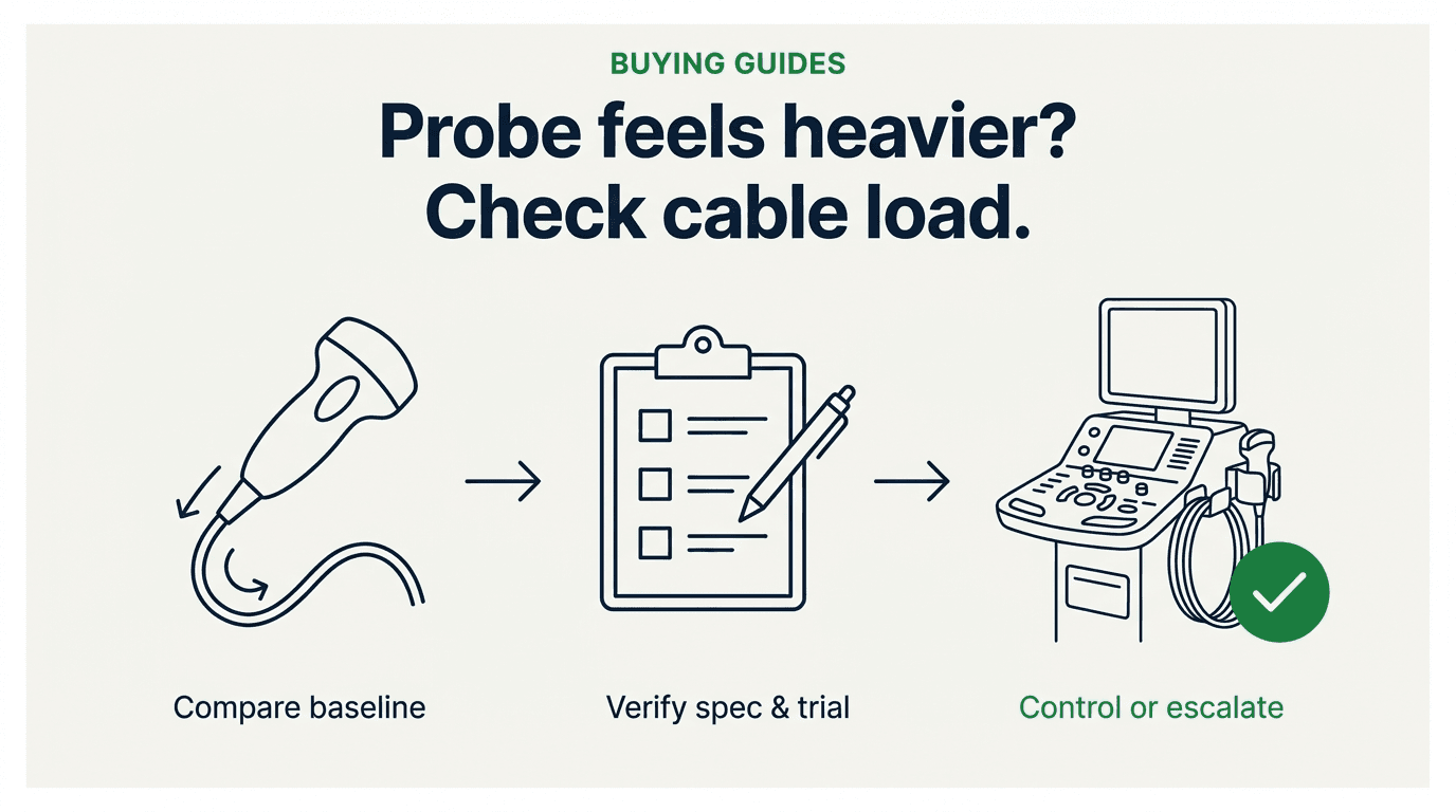
What Actually Changes When a Probe or Its Cable Is Replaced or Repaired?
Every hospital healthcare technology management (HTM) department, independent service organization (ISO), and imaging distributor already knows why a replacement or newly repaired transducer must pass physical, electrical, and acoustic gates before touching a patient. Our foundational ultrasound probe acceptance testing protocol covers those checks in three gates: Gate 1 physical inspection (including the acoustic lens under 10x magnification and the cable and strain relief), Gate 2 IEC 60601-1 / 60601-2-37 electrical-leakage testing, and Gate 3 in-air reverberation and tissue-equivalent phantom image checks. Those gates remain non-negotiable. To keep the two protocols distinct, this page labels its own checks Gates A–E.
What those three gates do not record is how the transducer loads the operator's hand, wrist, forearm, and shoulder during a clinical shift. In typical fleet operations—where probes face recurring service events across the replacement cycles examined in our ultrasound probe repair versus replace decision guide—a transducer returned from repair, re-cabling, or advance-exchange replacement can pass every electrical and acoustic test while differing mechanically from the unit that left the room:
- Cable bundle mass, jacket stiffness, and bend radius: A multi-element diagnostic array routes dozens to hundreds of micro-coaxial conductors plus shielding through a single jacket. If a re-cabling job substitutes a thicker aftermarket jacket, a stiffer outer braid, or a slightly shorter or longer cable assembly than the outgoing revision, the suspended loop between the console hanger and the patient bed pulls with a different downward and rotational force.
- Strain-relief geometry and cable-insertion lever arm: Where the cable enters the proximal end of the transducer handle, the molded strain relief sets the lever arm above the sonographer's grip. A stiffer or longer replacement strain relief shifts the effective center of mass away from the acoustic lens and increases rotational pull at the wrist when the probe is tilted.
- Re-housing shell balance and surface texture: A March 2025 article from repair and testing laboratory Acertara Acoustic Laboratories uses exactly this example: if a replacement housing 'alters weight distribution or grip texture, the probe’s ergonomic performance changes,' and existing QA protocols 'rarely capture or quantify these shifts' 6. A different shell material, wall thickness, or internal fill is the kind of change that can make a re-housed probe more front-heavy, back-heavy, or slicker in a gloved hand.
- Generational or revision differences across like-for-like replacements: Even when a replacement probe is an authentic, unmodified unit, OEMs revise cable materials across hardware revisions and platform generations. In a Toshiba-era ergonomics whitepaper (© 2012, hosted by Canon Medical Systems USA), ergonomist Dr. Richard Graveling of IOM Consulting noted that historical studies identified transducer cable weight and rigidity as contributors to musculoskeletal problems, while 'the use of lighter, more flexible cables in modern transducers helps to reduce these factors' 3. If an older-revision loaner or refurbished exchange replaces a newer low-drag revision—or if a department switches between compatible probe families—the operator feels the generational step immediately.
Because typical incoming QA records capture identity, leakage readings, and image results rather than handling characteristics, biomedical teams seldom have baseline notes on how the outgoing probe balanced in the hand or draped across the console holders. When sonographers report that the replacement 'feels heavier,' the complaint lands in a documentation vacuum.
What Do the Professional Standards Say About Cable Load, Balance, and Torque?
To build an acceptance check that neither invents fake specifications nor dismisses operator strain, clinical engineers must keep the authority hierarchy exact. Regulators, professional consensus bodies, OEM-hosted ergonomics authors, peer-reviewed researchers, and repair laboratories each address a different slice of the problem—and none of them publishes a numeric weight or torque cutoff.
At the regulator-adjacent layer, the US Occupational Safety and Health Administration (OSHA) Hospitals eTool — Clinical Services: Sonography 1 mentions the transducer cord in exactly one sentence: 'To scan a patient, they often must lean across the person’s body while holding a bulky transducer attached to a heavy cord.' OSHA notes that sonographers face musculoskeletal disorder (MSD) risk in their necks, shoulders, arms, and wrists, and cites the May 2003 SDMS industry standards and NIOSH Publication No. 2006-148 1. However, the scanning controls OSHA lists are workstation adjustability and work practice—height-adjustable exam tables, adjustable monitors and keyboards, keeping the wrist straight (bent less than 15 degrees), alternating right- and left-hand scanning where feasible, and anti-fatigue mats—while its remaining controls address patient handling and equipment transport 1. OSHA does not mention cable braces, cable holders, or transducer weight limits. Any vendor or internal policy claiming that 'OSHA requires cable braces' or 'OSHA sets probe weight limits' misstates the source.
The sonography-specific equipment and accessory recommendations live instead in the professional consensus standard: the Society of Diagnostic Medical Sonography (SDMS) Industry Standards for the Prevention of Work Related Musculoskeletal Disorders in Sonography, developed through a 2016 Consensus Conference of 26 organizations and published under a 2021 joint copyright 2. The SDMS document explicitly states in its background that its standards 'expand, and make specific to sonography, abatement methods included in the Occupational Safety and Health Act' 2. Its clauses use consensus 'should' and 'recommended' language across three sections directly relevant to transducer acceptance:
- Section I.A.4 — Manufacturer's Responsibility (Transducers): Clause (a) states that 'Transducers and cables should be lightweight and balanced to minimize torque on the wrist.' Clause (b) recommends designs that facilitate a palmar grip and a neutral wrist position. Clause (c) states that 'Transducer housing should be slip resistant and sized to fit the 5th to 95th percentile of the hand size of the user population to reduce grip force required to hold and manipulate the transducer.' Clause (d) recommends cables of suitable length for intended applications without interfering with equipment access or creating tripping or entanglement hazards. Clause (e) states that 'Low profile and easily accessible cable management systems are recommended,' and Clause (f) recommends easily accessible transducer holders capable of securing a variety of probes, including endocavitary probes 2.
- Section I.A.7 — Manufacturer's Responsibility (Accessories): Clause (b) recommends support devices for the scanning arm in abduction when applicable; Clause (c) states that 'A wearable transducer cable support device is recommended to allow the sonographer/user to reduce the grip by reducing the amount of torque on the wrist/forearm'; and Clause (d) recommends properly fitting, textured exam gloves to reduce grip force 2.
- Section II.A — Employer Responsibility (Risk Management): Clause 3 directs employers to conduct annual risk assessments in consultation with sonographers covering scanning workstation equipment, exam scheduling, and control measures. Crucially for procurement and incoming acceptance, Clause 4 states: 'Employer should conduct a needs assessment, with sonographer/user input, prior to the purchase of new equipment and maintain a budget that demonstrates ongoing planning toward adequate provision of ergonomic equipment and ancillary devices as part of the risk assessment process' 2.
By contrast, the American Institute of Ultrasound in Medicine (AIUM) Official Statement on Preventing Work-related Musculoskeletal Disorders (approved 2017; reapproved 2020 and June 15, 2026) establishes shared responsibility among employers, sonographers, and equipment manufacturers, calls for annual WRMSD education and microbreaks, and references the 2016 SDMS Industry Standards and the 2023 AIUM Practice Principles—but contains no cable-specific wording on its own official statement page 7. Table 1 maps what each source chain establishes and what none of them provides.
| Source & Document Era | Authority Type | What It Explicitly States on Transducer & Cable Load | What It Does Not Provide (Boundary) |
|---|---|---|---|
| OSHA Hospitals eTool: Sonography (current page; cites 2003 SDMS & 2006 NIOSH) [1] | US regulator educational eTool | Names leaning across patients while 'holding a bulky transducer attached to a heavy cord'; lists neck, shoulder, arm, and wrist MSD risk with workstation, work-practice, patient-handling, and transport controls. | Recommends no cable braces or holders; sets no weight, torque, or drag limit; not an enforceable transducer acceptance standard. |
| SDMS Industry Standards (2016 Consensus Conference of 26 organizations; PDF © 2021) [2] | Professional consensus standard | I.A.4: Transducers/cables 'should be lightweight and balanced to minimize torque on the wrist'; palmar grip; slip-resistant housing (5th–95th percentile hand size); low-profile cable management & accessible holders. I.A.7: Wearable cable support device to reduce grip by reducing wrist/forearm torque. II.A.4: Employer pre-purchase needs assessment with sonographer input. | Consensus 'should' / 'recommended' guidance—not a statutory mandate and contains no numeric gram, newton, or newton-meter pass/fail threshold. |
| Graveling R., IOM Consulting (Toshiba-era whitepaper, © 2012, Canon host) [3] | OEM-hosted independent ergonomics whitepaper | States studies identified 'transducer cable weight and rigidity as contributing to musculoskeletal problems' and lighter, more flexible modern cables help reduce them; explains tool-size-versus-hand-size grip strain, non-slip housings with a palm 'shoulder', and avoiding a 'pencil' grip. | Older (2012) console-framed whitepaper; provides technique and posture hierarchy rather than an incoming probe acceptance checklist. |
| Coffin CT & Baker JP (early-2000s Contemporary OB/GYN reprint, Canon host) [4] | OEM-hosted clinical-educator reprint | Explains that 'cable insertion into the transducer casing causes the transducer to be imbalanced and causes torque on the forearm'; notes that narrow casings require a tighter grip and very wide casings over-extend the fingers; warns that neck-draping and hip-trapping can injure the cervical or lumbar spine; endorses equipment holders or a below-elbow Velcro armband. | Carries 1997–1999 survey context; describes operator work practices and engineering-control concepts rather than post-repair QA records. |
| Zangiabadi et al., BMC Health Serv Res (2024; PROSPERO CRD42024507972) [5] | Peer-reviewed systematic review & meta-analysis | Pooled sonographer MSD prevalence 75.80% (95% CI: 65.37–86.23%, I² = 99.7%, 30 studies, n = 13,916); summarizes literature grip-force figures and identifies 'lightweight probes with cable holders' among ergonomic interventions. | Population-level epidemiological synthesis with extreme heterogeneity; does not link replacement probes to injury incidence or set per-device limits. |
| Acertara Acoustic Laboratories blog (18 March 2025) [6] | Repair-industry testing laboratory (competitor) | Notes QA protocols prioritize acoustic output, electrical safety, and mechanical integrity while rarely capturing post-repair ergonomic drift (re-housing weight distribution, grip texture, rigid-cable drag); proposes documented 'ergonomic equivalency'. | Advocacy position from a commercial testing lab; defines no step-by-step comparative bench protocol or public specification corpus. |
| AIUM Official Statement on Preventing WRMSDs (reapproved 15 June 2026) [7] | Professional society official statement | Establishes collaborative responsibility among employers, sonographers, and manufacturers; calls for annual WRMSD education, microbreaks, and safer equipment innovation. | Negative finding: contains no cable-specific language on its own statement page (cable-brace guidance lives in the SDMS standard). |
Source: Primary document synthesis across OSHA [1], SDMS (2016/2021) [2], Graveling (2012 Toshiba-era whitepaper) [3], Coffin & Baker (early-2000s reprint) [4], Zangiabadi et al. (2024 meta-analysis) [5], Acertara (March 2025) [6], and AIUM (2026 reapproved) [7].
Why Is 'Feels Heavier' an Acceptance Signal, Not Just a Comfort Complaint?
When a sonographer says a returned or replacement transducer 'feels heavier,' the physical difference on a scale may be small or even nonexistent. What the operator may be sensing instead is rotational torque at the wrist and forearm combined with the extra grip force needed to stop the probe from twisting or sliding on gel-covered skin.
The mechanical explanation is straightforward and has been documented for more than two decades. In their early-2000s clinical-educator article reprinted by Canon Medical Systems USA, Carolyn T. Coffin and Joan P. Baker explain: 'The cable insertion into the transducer casing causes the transducer to be imbalanced and causes torque on the forearm of the ultrasound practitioner' 4. A transducer does not hang vertically like a plumb bob; the sonographer holds it at an angle against the patient's abdomen, chest, neck, or extremity while the heavy multi-coaxial cable exits the top of the handle and loops across the bed or floor toward the console port. Whenever a replacement cable is stiffer, heavier per unit length, or routed with more unsupported drop, gravity and bending stiffness pull the tail of the probe sideways and downward. As Acertara's March 2025 analysis similarly notes, 'Cables that are too rigid create drag forces that alter the probe’s orientation' and front-heavy or unevenly weighted housings require compensatory grip adjustments 6.
To counter that rotational torque and prevent the acoustic lens from losing contact or rotating off-axis, the operator tightens their pinch or palmar grip. Casing geometry and surface finish govern how hard the hand must squeeze. Coffin and Baker note that narrow transducer casings require a tighter grip, while extremely wide casings over-extend the fingers 4. In the Toshiba-era ergonomics guide (© 2012), Graveling adds that non-slip housing material and a contoured 'shoulder' that sits in the palm help the operator use as little grip force as possible, that a 'pencil' grip is best avoided, and that a slipping probe should be wiped or held with a thin glove rather than gripped harder 3. Graveling also summarizes earlier exposure studies—citing Morton and Delf (2008) for reported pain and discomfort ranging from 63% to 89.7% across more than ten surveys, and Village and Trask (2007) for objective electromyography (EMG) evidence supporting the role of high grip force in hand and wrist problems alongside posture observations showing sonographers spending roughly 50% of scanning time with shoulders raised above 45 degrees and 40% with the neck bent or twisted over 20 degrees 3.
Current epidemiological synthesis confirms why incremental grip and torque load matters across a department's workday, even while requiring careful statistical interpretation. In a 2024 systematic review and meta-analysis published in BMC Health Services Research (PROSPERO CRD42024507972; searches through February 7, 2024 across 30 studies covering 13,916 sonographers), Zangiabadi et al. reported a pooled overall MSD prevalence of 75.80% (95% CI: 65.37%–86.23%), with extreme between-study heterogeneity (I² = 99.7%, P < 0.001; individual study prevalence estimates ranged from 20.23% to 99.3%) 5. By anatomical region, pooled estimates were 63.73% for the neck (95% CI: 56.17%–71.30%, I² = 98.9%), 60.13% for the shoulder, 53.69% for the upper back, 49.84% for the lower back, 44.41% for the wrist, and 27.46% for the elbow 5. Summarizing prior biomechanical literature in their introduction, the review authors note reported hand/wrist forces of at least 1 kg for 90% of scanning time, an average probe grip force of 3.96 kg across scanning sessions (rising up to 27.6 kg when scanning obese patients), and elevated WRMSD risk associated with a pinch-grip above 0.9 kg, a power-grip above 5.5 kg, or scanning more than four continuous hours per day 5.
Keep the boundary on these numbers clear: population prevalence and literature grip-force summaries describe the occupational baseline of sonography, not the failure rate of replacement probes. No study in the 2024 meta-analysis measures injury incidence from repaired or replacement transducers, and as we documented in our 4D motor board versus volume probe decision guide (which cites passive FDA MAUDE report 8020021-2016-00001, describing sonographer wrist and shoulder strain attributed to the weight of a volumetric probe, as an ergonomic incident rather than a motor fault), passive surveillance and population surveys do not prove per-device causation. What they do prove is why the SDMS Industry Standards treat lightweight, balanced transducers and cables, slip-resistant housings, cable management, and pre-purchase sonographer input as core equipment responsibilities 2.
How Do You Run a Comparative Cable-Load Check When No Numeric Threshold Exists?
Because none of these sources publishes a gram or newton-meter cutoff, weighing a replacement probe on a bench scale in isolation tells you nothing actionable. A 3D/4D mechanical volume probe is inherently heavier than a small-parts linear array; a phased-array cardiac cable carries a different coaxial bundle than a hockey-stick intraoperative probe. The only valid reference is comparative: how the incoming replacement or repaired transducer compares against (1) the OEM's documented model, part number, and cable specification, and (2) the outgoing probe of the same model—or a known-good sister probe in the same fleet—mounted on the same ultrasound cart.
This five-gate comparative check sits alongside—never in place of—the physical damage, IEC 60601-2-37 electrical leakage, and acoustic phantom gates defined as Gates 1–3 in our ultrasound probe acceptance testing protocol. Biomed runs Gate A at the incoming bench; Gates B and C are side-by-side comparisons on the actual console that biomed runs together with a lead sonographer; Gate D is the lead sonographer's exam-room trial; and Gate E closes the record before the service ticket or purchase acceptance is signed.
- Gate A — Identity, Revision, and Cable Specification Match: Compare both sides of the probe connector label, model designation, part number, serial number, connector housing style, and cable assembly markings against the purchase order, service work order, and OEM documentation. If the probe was sent out for repair or re-cabling, compare arrived photos against your pre-shipment intake photos. Record whether the cable jacket outer diameter, strain-relief boot length, and connector latch match the outgoing unit or sister probe.
- Gate B — In-Hand Housing Balance, Sizing, and Slip Resistance: Using the SDMS Section I.A.4(a)–(c) criteria as qualitative inspection prompts 2, hold the incoming transducer and a reference unit side-by-side in a palmar grip with a neutral wrist, both dry and with standard textured exam gloves (Section I.A.7(d)). Check whether a re-housed shell feels front-heavy or back-heavy compared to the reference unit, whether the housing seam or contour still supports a relaxed palmar grip without forcing a tight 'pencil' pinch 3, and whether the replacement housing material is unexpectedly slick 6.
- Gate C — Routed Cable Hang, Holder Fit, and Wrist Torque: Connect the probe to the actual ultrasound console and drape the cable through the system's low-profile cable management hooks and transducer holders (SDMS Section I.A.4(d)–(f)) 2. Verify that the nosecone seats securely in the console holder without popping out, that the cable length reaches the patient side of the table without pulling taut or dragging excess coils on the floor (a tripping and caster-crush hazard), and whether the suspended cable loop twists the probe handle in the operator's relaxed palm.
- Gate D — Task-Specific Seated and Standing Scanning Trial: Before final sign-off, have a sonographer who regularly performs the room's longest or most demanding studies evaluate the probe in the actual exam room configuration (described in detail in the next section).
- Gate E — Acceptance Record & Baseline Archiving: Record the observer names (biomed engineer and lead sonographer), the reference probe serial number used for comparison, photographs of the connector labels and console cable routing, and the routing disposition (accepted as equivalent, accepted with workplace cable-support controls, or held as a supplier deviation).
| Comparative Gate | What to Compare (Reference vs. Incoming) | Standards / Literature Basis | Synthetic Illustration: Pass vs. Deviation Record | Disposition / Next Action |
|---|---|---|---|---|
| Gate A: Identity & Cable Spec Match | Model, part number, connector label (both sides), strain-relief boot geometry, cable jacket OD and length against outgoing unit or sister probe. | SDMS I.A.4(d), (g) (suitable cable length; single-handed minimal-force connector) [2]; Acertara post-repair QA record gap [6]. | Synthetic Example: Outgoing probe had flexible 10.5 mm OD cable and 55 mm strain relief; returned re-cabled unit arrived with stiff 12.8 mm OD generic jacket and 85 mm rigid boot not disclosed on quote. | Hold sign-off; open documented supplier deviation with side-by-side photos. |
| Gate B: In-Hand Balance & Palmar Grip | Center-of-gravity feel in palmar grip, housing contour/shoulder, and surface slip resistance with textured exam gloves. | SDMS I.A.4(a)–(c) (lightweight/balanced; palmar grip; slip-resistant 5th–95th percentile housing) & I.A.7(d) [2]; Graveling [3]; Acertara [6]. | Synthetic Example: Re-housed convex probe shell has glossy smooth finish lacking the original matte texture, requiring visibly tighter pinch grip when gel is present. | Quarantine for supplier housing correction; do not release slick re-housed shell. |
| Gate C: Routed Cable Hang & Holder Fit | Probe seating in console cup; cable routing through cart hooks; rotational twist on wrist at primary reach zone; floor clearance. | SDMS I.A.4(a), (d)–(f) (minimize wrist torque; no tripping/entanglement; low-profile cable management & accessible holders) [2]; Coffin & Baker [4]. | Synthetic Example: OEM replacement revision matches part number, seats in holder, and clears floor, but slightly stiffer new jacket adds minor pull when reaching across exam table. | Pass hardware spec; route to Gate D trial with console boom/holder and below-elbow cable brace. |
| Gate D: Task-Specific Clinical Trial | Seated and standing reach across exam table in the department's actual protocol without triggering neck-draping or hip-trapping workarounds. | SDMS II.A.4 (sonographer input) & III.A (neutral posture) [2]; Coffin & Baker (hazardous workarounds) [4]; Graveling (arm angle <45°) [3]. | Synthetic Example: Lead sonographer completes simulated bilateral lower-extremity venous positioning; with cable clipped to cart holder and forearm armband, wrist stays neutral (<15°). | Release to clinical service with documented holder/armband setup in room checklist. |
| Gate E: CMMS & Service Record Closure | Observer sign-off (HTM + sonography), comparator serial number, intake vs. return photos, and final disposition. | Acertara post-repair ergonomic record proposal [6]; Rongtao intake/QC photo documentation workflow [8]. | Synthetic Example: CMMS ticket logs comparator S/N, routing photos, and sonographer co-signature before warranty acceptance window closes. | Archive as baseline for future repairs or replacements of that probe asset. |
Source: Rongtao Medical acceptance synthesis grounded in SDMS Sections I.A.4, I.A.7, and II.A.4 [2], Graveling [3], Coffin & Baker [4], and Acertara [6]. All illustrative record values are synthetic examples—not universal pass/fail limits.
The Task-Specific Trial: Grip, Drag, and Reach Before Sign-Off
A thirty-second bench check in the biomed shop cannot replicate how a transducer cable behaves across an examination table. In the biomed shop, the engineer typically holds the probe eighteen inches from the console port with the cable hanging slack. In a clinical exam room, the sonographer must reach the far side of the patient's body while keeping the scanning arm near neutral abduction, viewing the monitor without cervical twisting, and maintaining steady acoustic coupling pressure.
As Graveling's ergonomics whitepaper summarizes from Burnett and Campbell-Kyureghyan (2010), different ultrasound procedures load the operator differently: average transducer force was much higher in deep vein thrombosis (DVT) studies, while average wrist deviation was much higher in thyroid examinations 3. Similarly, the literature summarized in the 2024 meta-analysis by Zangiabadi et al. reports that grip force can rise sharply when scanning obese patients and links more than four continuous hours of scanning per day to elevated WRMSD risk 5. For that reason, Gate D should match the probe's actual clinical assignment rather than a generic phantom swipe:
- Set the workstation to neutral baseline first: Before judging the probe, adjust the exam table height, operator chair, monitor height, and console position so the sonographer works close to neutral: OSHA describes a straight wrist (bent less than 15 degrees) with the elbow close to the body 1; SDMS calls for table height that keeps scanning-arm abduction under 30 degrees and recommends keeping 80% of work within the primary reach zone 2; and Graveling notes that raising the arm beyond about 45 degrees can restrict shoulder tendon blood flow 3. Otherwise, a poorly positioned cart or fixed-height bed will make any cable feel like it is dragging.
- Compare far-side reach in both seated and standing postures: Position the transducer at the furthest anatomical reach required by the room's standard protocol (for example, left-sided cardiac apical window, contralateral lower-extremity venous compression, or obstetric lateral sweep) with the cable routed through the console's overhead or side cable hook. Check whether the cable pulls taut, lifts out of the holder, or forces the wrist into ulnar/radial deviation.
- Compare palmar grip stability under gel contact: With standard textured exam gloves (SDMS Section I.A.7(d)) 2, verify that the sonographer can rotate and angle the probe using whole-forearm movement rather than finger-pinch twisting 3.
- Test with the room's wearable cable support device attached: If the department uses a below-elbow Velcro cable armband or wearable support device (SDMS Section I.A.7(c); Coffin & Baker) 2 4, verify that the replacement cable diameter fits the clip or strap cleanly without slipping or kinking the jacket.
Synthetic Acceptance Trial Illustrations (Case A vs. Case B)
The two scenarios below are synthetic operational illustrations constructed to show how the comparative record separates a supplier defect from a workplace-routing fix. They do not represent real patient exams, staff records, or universal weight limits.
Synthetic Case A (Supplier Deviation — Re-Cabled Vascular Linear Probe): A hospital HTM team receives a repaired high-frequency linear probe used for lower-extremity DVT studies. Gates 1–3 (physical damage, IEC 60601-2-37 leakage, in-air reverberation, and phantom QA) pass. However, during the Gate A/C/D comparison against an identical sister probe on the same cart, biomed and the lead vascular sonographer document that the replacement cable jacket is visibly thicker and stiffer than the original OEM cable, refuses to stay seated in the console's low-profile side clip, and twists the probe handle 30 degrees off-axis whenever the operator relaxes their fingers. Decision: Quarantine and escalate to the repair vendor with side-by-side photographs of the sister probe's cable specification and holder seating.
Synthetic Case B (Design Trajectory — Refurbished Abdominal Convex Replacement): An outpatient clinic replaces a failed abdominal convex transducer with an authentic, earlier-revision OEM unit of the same model family. Gate A confirms exact OEM part-number and connector compatibility. During Gate D, sonographers note the older-revision cable is slightly less supple than the failed late-revision unit, causing mild drag during standing right-upper-quadrant sweeps when the cord hangs free to the floor. When routed through the console's upper cable management arm and paired with a below-elbow Velcro cable support band, wrist torque drops to match the room's baseline feel. Decision: Accept the probe, equip the room with the wearable support band, and update the room setup card.— Synthetic Comparative Acceptance Trial Examples
Workarounds That Should Fail Review: Neck Draping and Hip Trapping
One of the clearest indicators that a replacement probe's cable load or routing has not been properly managed is how sonographers improvise when left with a dragging cord. If an acceptance trial or post-deployment room audit reveals staff using their own bodies as cable anchors, the configuration should fail review immediately.
Coffin and Baker state the mechanism and the injury hazard explicitly in their clinical-educator reprint: 'The cable insertion into the transducer casing causes the transducer to be imbalanced and causes torque on the forearm of the ultrasound practitioner. To compensate for this, practitioners may either drape the cable around their necks or, if they are standing to scan, trap the cable between their hips and the exam table. Both of these work practices can produce injuries of the cervical or lumbar spine. The cable should be draped in holders on the ultrasound equipment or managed with an armband that is worn below the worker’s elbow and entraps the cable in a Velcro® tab' 4.
Why do these two workarounds appear so often after a probe replacement or room change, and why must both biomed and sonography leadership treat them as red flags?
- Draping the transducer cable around the neck: Looping the cord over the cervical spine unloads the downward pull from the scanning wrist, which immediately makes a heavy or stiff cable feel lighter in the hand. However, it transfers the static load and every tug of the cable directly onto the cervical vertebrae and trapezius muscles—the exact anatomical region that already carries the highest pooled MSD prevalence in the 2024 meta-analysis (63.73%, 95% CI: 56.17%–71.30%, I² = 98.9%) 5, while tying the operator to the cable if the patient moves or the cart rolls.
- Trapping the cable between the hips/pelvis and the examination table: When standing to scan, pinning the cord against the table rail with the hip stops the cable loop from sliding back toward the console. Doing so locks the operator's pelvis and lumbar spine into an asymmetric, twisted static posture while the upper torso reaches across the patient—the practice Coffin and Baker link to lumbar spine injury 4—and it repeatedly pinches the cable jacket against the table edge.
During Gate D of acceptance, if the only way a sonographer can keep a newly repaired or replacement probe comfortable is to drape the cord over their shoulder/neck or pin it with their hip, do not sign off the room as ready. Either the probe has a cable-specification/stiffness defect that belongs back with the supplier, or the workstation lacks the accessible cable hooks and wearable below-elbow support recommended by SDMS Sections I.A.4(e) and I.A.7(c) 2 4.
When Is a Heavier Probe a Defect Versus a Design Difference?
A disciplined acceptance process protects both the healthcare facility and the service provider from guesswork. Not every heavier-feeling probe is defective, and not every complaint can be solved by handing the sonographer a Velcro strap. The comparative evidence gathered in Gates A through D determines whether the observation routes back to the vendor as a technical deviation or stays in-house as an ergonomic workplace-control adjustment.
When evaluating whether to escalate to an OEM or independent repair supplier—a distinction explored in our guide to OEM versus independent probe repair and our checklist to qualify an ultrasound probe repair provider—use the evidence-gated criteria in Table 3.
| Observed Condition During Gates A–D | Root-Cause Classification | Primary Evidence Required | Responsible Owner | Action & Escalation Path |
|---|---|---|---|---|
| Replacement cable after repair/re-cabling has visibly larger diameter, stiffer bend radius, or heavier drag than a sister probe of the exact same part number. | Supplier-escalable defect (non-equivalent cable assembly). | Side-by-side photos of incoming vs. sister probe cable markings/OD, strain relief, and holder hang; work-order scope review. | Repair vendor / refurbisher + HTM biomedical engineering. | Quarantine probe; submit documented deviation pack to repair provider to re-cable with matching specification or exchange. |
| Re-housed transducer shell alters weight distribution (front/back-heavy) or replaces a contoured matte grip with a slick or poorly fitting shell. | Supplier-escalable defect (post-repair rehousing drift) [6]. | Intake vs. post-repair housing photographs; side-by-side palmar grip comparison with reference unit. | Repair vendor / refurbisher + HTM biomedical engineering. | Hold sign-off; return under repair warranty for OEM-equivalent housing geometry and slip-resistant finish. |
| Strain relief or cable jacket has internal kinking, twist memory, or binding at the handle entry that torques the wrist when rotated. | Supplier-escalable mechanical defect. | Video or photo showing cable twist/kink when draped on console holder; Gate 1 physical inspection record. | Supplier / repair vendor. | Reject at Gate 1 / Gate C; return for strain-relief and cable-termination rework before clinical release. |
| Authentic replacement probe matches OEM part number and spec, but is an older hardware revision than a newer 'lightweight/flexible cable' revision. | Legitimate design difference (generational OEM trajectory) [3]. | Both sides of connector label confirming authentic OEM part/revision; clean Gates 1–3 QA record. | Clinical department + HTM / workplace ergonomics. | Accept hardware; deploy SDMS-endorsed console cable holders and wearable below-elbow cable support [2, 4]; log revision note in CMMS. |
| Specialty probe (e.g., mechanical 3D/4D volume probe or matrix array) feels heavier than a standard 2D curved array. | Inherent transducer architecture difference (motor/array mass). | Model specification confirmation (see our 4D motor-board vs. volume-probe guide). | Clinical department + employer risk management (SDMS II.A) [2]. | Do not route as a probe defect; apply task rotation, exam scheduling limits, arm abduction supports, and cable holders [2, 4]. |
| Probe and cable match reference unit on the bench, but drag in the exam room because the console holder is broken or the table/cart layout forces a long reach. | Workstation / environmental control gap. | Exam room layout audit showing missing/broken cable hook or excessive reach outside primary reach zone [1, 2]. | Employer / facility engineering & sonography management. | Replace broken console cable holders (SDMS I.A.4(e)–(f)) and reposition cart/table per OSHA and SDMS neutral-posture controls [1, 2]. |
Source: Rongtao Medical synthesis based on SDMS Sections I.A.4, I.A.7, and II.A.4 [2], Graveling [3], Coffin & Baker [4], and Acertara [6].
After Acceptance: What Do Holders and Supports Do — And Not Do?
When Gate A through Gate D confirm that an incoming probe matches its OEM specification and has no repair-induced rehousing or re-cabling defect, managing its operational load moves from supplier QA to workplace engineering and administrative controls. Here again, accuracy about what the published evidence supports—and what it warns against—prevents false security.
| Practice or Device | Status in Published Standards & Literature | Biomechanical Purpose | Documented Limits & Counter-Evidence (What It Cannot Do) |
|---|---|---|---|
| Low-profile console cable management hooks & accessible probe holders | Recommended — SDMS Section I.A.4(e)–(f) [2]; Coffin & Baker [4]; Zangiabadi et al. [5]. | Carries suspended cable weight on the ultrasound cart rather than the operator's wrist; prevents floor tripping and caster entanglement. | Only relieves cable loop weight between the cart and bed; does not correct excessive arm abduction (>30°–45°) if the patient is positioned too far away [3, 4]. |
| Wearable transducer cable support device / below-elbow Velcro armband | Recommended — SDMS Section I.A.7(c) [2]; Coffin & Baker [4]. | Anchors the cable just below the scanning elbow so rotational torque is taken off the wrist and hand grip force can relax [2, 4]. | Transfers a portion of cable weight to the forearm/arm; no source guarantees injury prevention, and it cannot compensate for a non-conforming stiff aftermarket cable. |
| Properly fitting, textured exam gloves | Recommended — SDMS Section I.A.7(d) [2]. | Increases friction against the transducer housing so the operator needs less pinch/squeeze force when gel is present. | Mitigates surface slip only; does not alter transducer mass or cable-insertion torque. |
| Arm abduction support devices / cushions | Recommended when applicable — SDMS Section I.A.7(b) [2]; Coffin & Baker [4]; Graveling [3]. | Supports the weight of the scanning arm when abduction exceeds neutral posture. | Graveling explicitly warns arm rests are 'beneficial in the short term... no substitute for working on getting the arm angle down' (<45° shoulder abduction) [3]. |
| Height-adjustable exam tables, monitors, keyboards & alternating hands/task rotation | Recommended — OSHA eTool [1]; SDMS Sections I–III [2]; AIUM [7]. | Keeps most work within the primary reach zone (SDMS recommends 80% of work there, elbows at the sides) and supports muscle-recovery microbreaks. | Requires active room setup before each exam and employer scheduling discipline (SDMS II.A & III.A) [2]. |
| Draping cable around the neck OR trapping cable between hips and exam table | Documented hazard — Reject practice (Coffin & Baker) [4]. | Improvised attempt by operator to unload wrist torque or stop cable slide. | Coffin & Baker warn that both practices 'can produce injuries of the cervical or lumbar spine' [4]; signals missing holders or excessive cable drag. |
Source: Synthesis of OSHA [1], SDMS Industry Standards Sections I.A.4, I.A.7, II.A, and III.A [2], Graveling [3], Coffin & Baker [4], and Zangiabadi et al. [5].
Counter-evidence and methodological boundaries to keep in view:
- No device guarantees injury prevention, and posture comes first: While SDMS Section I.A.7(c) recommends wearable cable support devices to reduce wrist/forearm torque 2, no published standard or study claims that a cable brace, holder, or lightweight cord guarantees prevention of WRMSDs. Graveling's ergonomics guide explicitly notes that arm supports 'might be beneficial in the short term, especially for those with existing shoulder problems, [but] they are no substitute for working on getting the arm angle down' (noting that raising the arm above 45 degrees restricts shoulder tendon blood flow and above 60 degrees risks brachial plexus tension) 3.
- Subjective feel alone does not prove a defective repair: Because OEMs actively engineer lighter, more flexible cables in newer models 3, a like-for-like replacement of an older model or a switch between transducer families can feel heavier without any defect in materials or workmanship. Only a comparative check against the outgoing unit or sister probe and OEM documentation separates true nonconformity from perception.
- Document-era and source-independence limits: The clearest published statements of cable weight and rigidity (Graveling © 2012 3) and of cable-insertion torque and neck/hip draping hazards (Coffin & Baker's early-2000s reprint citing 1997–1999 surveys 4) are older documents hosted on an OEM download portal. Their biomechanical descriptions remain valid, but current quantitative context should rely on the 2016/2021 SDMS consensus standards 2 and the 2024 meta-analysis 5—remembering that the meta-analysis itself carries very high heterogeneity (I² = 99.7%) and relies on self-reported questionnaires across cross-sectional studies. Furthermore, Acertara's March 2025 article 6 is cited solely as a repair-industry laboratory's public recognition that existing QA protocols rarely record post-repair ergonomic changes.
Where Rongtao Medical Fits — And Where It Does Not
Rongtao Medical is an independent ultrasound repair and parts organization serving hospital HTM departments, independent service organizations, refurbishers, and medical equipment distributors. It is not an occupational-health clinic, an ergonomics consultancy, a regulatory body, or an OEM's authorized representative.
Where Rongtao Medical fits:
- Probe and transducer repair & replacement sourcing: Fault diagnosis and element-level repair of linear, convex, phased-array, endocavitary, TEE, and volumetric (4D) probes through our Rongtao probe repair services, with turnaround and scope quoted per probe 8.
- Documented intake, QC, and packaging evidence trail: Our documented workshop workflow captures photo evidence at intake, quality control, and packaging, alongside a typical 90-day warranty and lifelong consultation on repaired items 8. For ultrasound circuit boards, every repaired board runs a 48-hour real-machine test inside an actual ultrasound system with a standard 5–8 business-day turnaround 8.
- Actionable deviation review: Because that workflow photographs items at intake, QC, and packaging, a customer who documents a post-repair or replacement discrepancy in connector labeling, strain-relief geometry, housing finish, or cable assembly against the outgoing unit has a concrete record to reopen with our engineering team.
Where Rongtao Medical does not fit:
- No claimed quantified ergonomic-equivalency certification: Industry commentary notes that existing QA protocols rarely quantify post-repair ergonomic change 6, and Rongtao does not claim to perform laboratory gram- or newton-meter ergonomic equivalency certification. Our verification is identity, physical inspection, and functional testing, with photo evidence on file 8.
- Not workplace ergonomics or medical advice: Room layout, exam scheduling, wearable cable braces, and occupational health assessments belong to the employer, clinical leadership, and qualified ergonomics specialists under SDMS and AIUM guidance 2 7.
Quote-ready handoff for a probe repair, replacement, or deviation review: When requesting a probe repair quote or escalating a comparative cable-load discrepancy on a received transducer, send the following evidence pack through our contact page so our engineers can verify the exact assembly immediately:
- Ultrasound console: OEM brand, system model, and software/hardware revision.
- Transducer identity: Exact probe model and OEM part number, plus clear photographs of both sides of the connector label and serial number tag.
- Comparative observation (for a heavier/dragging replacement): Side-by-side photographs of the incoming probe and the outgoing or sister reference probe showing the cable jacket markings, outer diameter, strain-relief boot, housing finish, and how the probe seats in the console holder.
- Electrical & image QA status: Summary of Gate 1–3 acceptance results (visual inspection, leakage test, in-air reverberation / phantom status).
- Commercial request: Quantity, desired path (repair of existing unit, re-cable specification check, or replacement/exchange quote), and shipping destination.
Frequently Asked Questions
Q: Our replacement probe passed electrical and image acceptance, but staff say it scans heavier - do we have to accept it?
A: Electrical leakage and phantom image quality are necessary gates, not the whole acceptance decision. Before final sign-off, run the five-gate comparative check against the outgoing probe or a sister unit of the same model. If the heavier feel comes from a supplier-escalable defect—such as a stiffer non-equivalent replacement cable, an altered strain-relief lever arm, or a slick or unbalanced re-housed shell—hold acceptance and escalate to the vendor with side-by-side photographic evidence. If the unit matches the authentic OEM specification and the difference reflects a hardware revision or probe family change, accept the hardware and address the load with console cable holders and a wearable below-elbow support.
Q: Is there a published weight or torque limit for transducer cables?
A: Not in any source reviewed for this guide. Neither OSHA's Hospital eTool 1, the SDMS Industry Standards 2, nor the AIUM official statement 7 publishes a numeric gram, newton, or newton-meter acceptance limit for ultrasound probes or cables. SDMS Section I.A.4(a) uses qualitative consensus language—'Transducers and cables should be lightweight and balanced to minimize torque on the wrist' 2. Because no universal numeric threshold exists, acceptance checks must be comparative against the outgoing unit or OEM documentation rather than based on an invented weight cutoff.
Q: The old probe's cable draped fine; the new one drags - is that the cable or something else?
A: Check three possibilities in order. First, compare the cable itself (Gate A and Gate C): a re-cabled probe or different hardware revision may have a stiffer outer jacket, a different cable length, or twist memory that fights the console hooks. Second, inspect the workstation holders: a missing, bent, or poorly positioned console cable hook forces a longer unsupported cable drop to the floor. Third, verify exam room geometry: if the table or cart was moved so the sonographer must reach further across the patient, any cable will pull harder against the wrist.
Q: Are cable braces or armbands an acceptable fix for a heavy cable?
A: For a probe that meets its authentic OEM specification, yes—as a workplace mitigation, not a cure-all. SDMS Section I.A.7(c) explicitly recommends 'a wearable transducer cable support device... to allow the sonographer/user to reduce the grip by reducing the amount of torque on the wrist/forearm' 2, and Coffin and Baker endorse managing the cord with equipment holders or an armband worn below the elbow 4. However, a wearable brace should never be used to excuse a defective aftermarket re-cabling job, and no published source guarantees that a brace alone prevents musculoskeletal injury.
Q: Should we put the probe and cable on a scale as part of acceptance?
A: A scale reading by itself is meaningless because there is no published pass/fail weight table and because total mass on a bench scale does not measure how the suspended cable torques the wrist when plugged into the cart. If your department chooses to record head-and-cable mass on a scale, do so only as a relative baseline log comparing an outgoing probe before repair against the same unit upon return—paired with in-hand balance and routed-holder checks.
Q: Who owns this check - biomed, sonography, or the supplier?
A: It is a shared handoff across all three. Biomedical engineering owns Gate A (verifying part numbers, connector labels, and intake-versus-return hardware match) and the CMMS acceptance record (Gate E). Gates B and C are joint biomed–sonography comparisons on the actual console, and sonography leadership owns the task-specific room trial (Gate D), aligning with SDMS Section II.A.4's recommendation for sonographer input prior to purchasing new equipment 2. The supplier or repair vendor owns delivering an assembly that matches the quoted specification and resolving any documented physical or cable-specification mismatch.
Q: Can we ask the repair vendor to document post-repair weight and balance against the original?
A: Yes, and doing so strengthens provider qualification. While existing QA protocols rarely capture or quantify post-repair ergonomic change 6, you can require your repair provider to capture intake and pre-shipment photographs of the housing, strain relief, cable markings, and connector labels, and to disclose in writing before quoting whether a repair involves re-housing or cable assembly replacement.